


: The Weight Loss Injection Revolution")


Globally, technological innovation is advancing at a breakneck pace, and the contemporary developments in the field of medicine are unprecedented. The ability to seamlessly treat diseases depends on time, and the sooner an illness is detected, the earlier it can be treated. Slowdowns in diagnosis may negatively impact patients’ prognoses.

Engineers and scientists are developing revolutionary tools and methods to diagnose and manage ailments swiftly and with exceptionally small sample sizes. Among the tools that help improve disease detection are:
Microscopes
Microscopes symbolize medical advancement since they offer a crucial viewpoint in scientific studies, deliver accuracy during diagnostic and surgical operations, and improve clinical results. They assist in providing precise diagnostics, resolving challenging research problems, and enhancing patient treatment outcomes. Medical professionals can observe and examine things ordinarily invisible to the naked eye using microscopes to investigate components at the micron scale.
Despite some viewing electron microscopy (EM) as outdated, it remains the gold standard for investigating infectious ultrastructure, pathogenesis, and clinical viral diagnosis. EM is particularly advantageous for tracking new illnesses and suspected viruses in the medical environment.
Identifying the pathogenic agent without needing organism-specific reagents is one of the essential benefits of employing EM for viral diagnosis. Even now, in the era of molecular diagnostics, EM remains a crucial tool for identifying novel and uncommon epidemics. Luckily, there are various options for microscopes to choose from.
Ultrasound
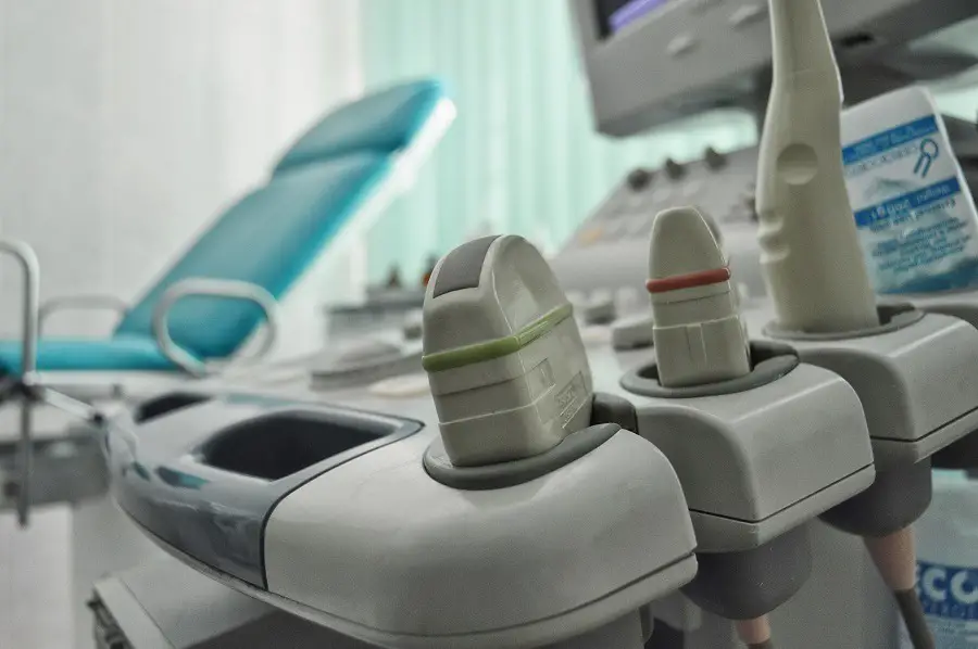
Ultrasound is a medical imaging technique that utilizes high-frequency sound waves to capture images of the body’s internal structures in real-time. This noninvasive test is commonly used to scrutinize internal organs, detect abnormalities in tissues and vasculature, and monitor fetal development during pregnancy. This noninvasive routine uses an instrument called a probe that is moved over the skin’s surface.
To obtain higher-quality images, a gel is applied to the skin. Throughout the process, an echo generated by the sound waves is converted into several explicit photos displayed on the screen. It is crucial to note that, unlike a CT scan or X-ray, this examination does not involve ionizing radiation. Instead, a computer interprets the waves to create an image to be used for diagnosis.
Biopsy
A biopsy entails the removal of sample tissues or cells for evaluation to ascertain the existence or severity of a medical condition. There are several types of biopsies, including bone marrow, endoscopic, needle, cutaneous, and surgical, and your physician will determine which one is necessary for your particular case. After the cells are successfully removed, a laboratory technician analyzes them.
Biopsies are also carried out to identify additional reasons for your symptoms, such as immune illnesses like chronic pancreatitis, inflammatory disorders like those in the kidney or liver, infections like tuberculosis, and other infections in general. A biopsy is also performed to determine patient compatibility for an organ transplant to ensure that the patient’s body will not reject it.
Magnetic Resonance Imaging (MRI)
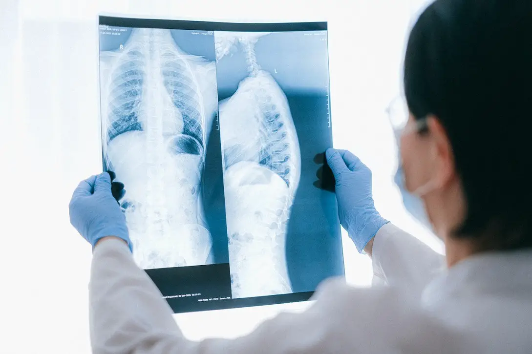
Magnetic resonance imaging (MRI) uses a powerful magnetic field and radio waves from a computer to provide precise images of the tissues and organs inside the body. An MRI is considerably more detailed than an X-ray in that it displays the patient’s muscles, nerves, bones, and other organs.
The patient lies inside the MRI scanner, a sizable tube filled with strong magnets. The magnetic field of MRI equipment causes the water molecules in the patient’s body to rearrange momentarily. When exposed to radio waves, these aligned atoms emit tiny signals, which are then used to form cross-sectional MRI images resembling bread slices. Since it creates detailed pictures of the inside of the body, it aids in diagnosing many conditions. A doctor can evaluate a patient’s organs, skeletal system, and tissues without causing any harm by using an MRI.
Computer Axial Tomography (CT or CAT) Scan
A CAT scan creates several cross-sectional images of particular sections of the examined object enabling medical professionals to view the thing without cutting it. The examination helps medical professionals detect clots in the blood, infections, tumors, and other illnesses. The X-rays swirl around a patient’s entire body as the scanner’s table goes slowly through the scanner. A buzzing or whirring sound is typical, and patients are required to remain completely still during the procedure to prevent any movement that could distort the image.
Positron Emission Tomography (PET) Scan
PET scans use a nuclear medicine available imaging technology to monitor metabolic processes in the human body, including blood flow, oxygen utilization, and sugar metabolism. A small quantity of radioactive glucose is administered into the patient’s blood vessel. A scanner then creates intricate computerized images of the body’s glucose-absorbing organs. The images can be used to find cancerous cells in the body’s tissues because they frequently absorb more glucose than healthy cells.
Final Thoughts
Engineers excel at finding technical solutions, while doctors require quick, accurate tools to diagnose diseases. A doctor’s ability to make the best treatment decisions will be aided by such precision, which might mean the difference between health and illness.
")


