Overview & Description
A breast ultrasound is a test that uses high-frequency sound waves toform images of tissues and other structures inside the breast.
Who is a candidate for the test?
Doctors may recommend this test so that they can:
An ultrasound may also be used to evaluate a woman who has possible signs ofbreast cancer.In some cases, this test is used instead of a mammogram.Some examples of when this test might be used include:
How is the test performed?
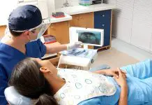
The test takes about 15 minutes. A healthcare provider can perform thistest in an office, clinic, or hospital. Usually, a woman puts on a hospital gown thatopens at the front before the test.
There are two ways to perform the test.
In either method, the sound waves bounce off internal tissues of the breastand then return to the scanning tool. A computer converts the sound waves into ablack-and-white image. The healthcare provider can then read this image of the internalpart of the breast.
In some cases at the time of the ultrasound, a doctor may insert a needleinto the breast to obtain tissue for a breast biopsy.The images from the ultrasound help guide the needle into the right area of the breast.
When the test is finished, the healthcare provider will dry the breast or wipe the gel off. Thewoman may then dress and leave.
Preparation & Expectations
What is involved in preparation for the test?
On the day of the test, the woman should not put any lotions or powderson her breast. No other preparation is generally required.
Results and Values
What do the test results mean?
Test results are usually sent to the woman’s healthcare provider, whothen discusses them with her. In some cases, ultrasound will show no problem in thebreast. Abnormal findings may include:
Article type: xmedgeneral


